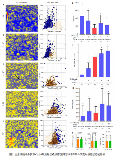
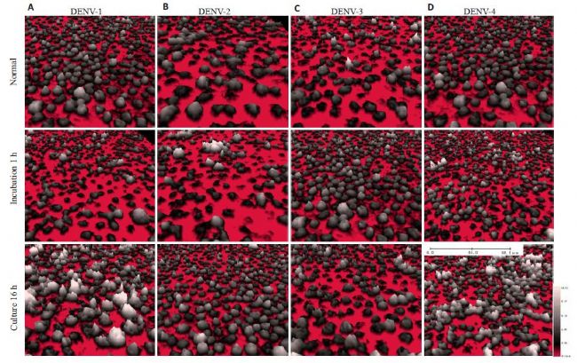
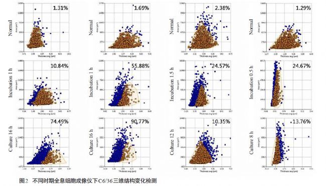
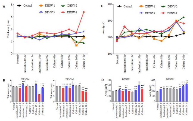
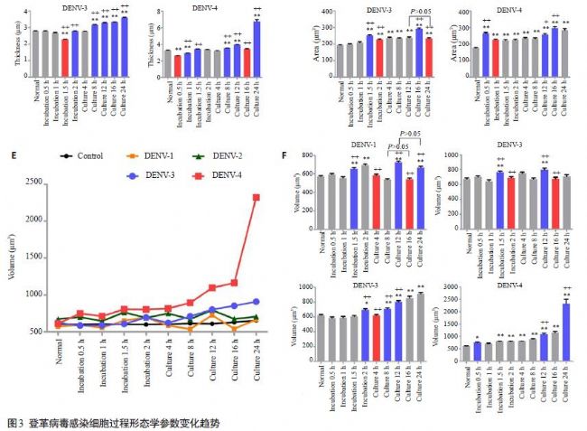
3D Morphology of Dengue Virus Infected C6/36 Cells Based on Digital Holographic Microscopy Digital holographic microscopy is a new technology that has been applied to the study of living cell morphology in recent years. The combination of digital holography and microscopy is used to specifically measure cell morphology evaluation parameters such as cell number, cell area, thickness and volume, and provide spatial, temporal, and high-resolution three-dimensional morphological images, which are transformed by data processing. It is a cellular change process such as cell proliferation, migration, activity and cell death [13-16]. Digital holographic microscopy has been gradually applied to the study of cell-level mechanisms, such as real-time imaging technology to study the pathophysiological changes of cells at various stages from principle to application [17]. At present, the mechanism of dengue virus infection is mainly through the research and expression of related proteins, cell pathways, cytokines, etc. However, there is no effective research method for the conformational change form and direction of virus-cell membrane fusion during invasion. Therefore, this study used the HoloMonitor M4 holographic cell imaging and analysis system to monitor the morphological changes of dengue virus-infected host cells in real time, and quantitatively analyze the changes of three-dimensional parameters such as cell area, thickness and volume during viral infection. Helps clarify the details of dengue virus-infected cells. At the same time, this method is equally applicable to other viral infection mechanisms, providing a new direction for studying cell morphological changes during viral infection. results and analysis 2.1 Optimal imaging density under C6/36 cell holographic cell imager Five different conditions, such as 10 5 , 2 × 10 5 , 4 × 10 5 , 8 × 10 5 , 10 6 / well , were established in the amount of cells to be inoculated. Three-dimensional morphological observation and cells were observed after overnight culture in each experimental group. Parametric analysis was performed to determine the optimal density of three-dimensional morphological experiments on dengue virus-infected C6/36 cells. The results showed that when the cell inoculation amount was 4×10⁵ in experiment 3, the living space of single cells was sufficient after adherence of C6/36 cells, showing a good circular three-dimensional shape, and the morphological characteristics of cells were consistent, suggesting that the three-dimensional imaging of cells has a high uniformity. Sex. At the same time, in the statistical analysis, whether it is cell area, thickness or volume, the standard deviation of the experimental group 3 data is the smallest, indicating that the degree of dispersion is the smallest under the inoculum, and each index has stronger uniformity. 2.2 Three-dimensional morphological changes of dengue virus-infected C6/36 cells At 4×10 5 inoculation, the C6/36 cell hologram showed that the three-dimensional morphology of single cells was uniform, and the cell area, thickness and volume dispersion were the smallest (P<0.05), which was determined to be the best imaging density; transfer to 37 °C There was no significant difference in cell thickness, area and volume after 5% CO 2 culture for 24 h (P>0.05); 4 serotypes of DENV group during incubation and culture The cell area and volume increase were consistent, but DENV1-4 showed different cell thickness reduction during incubation. The thickness of DENV1 and 2 cells decreased while DENV3 and 4 showed opposite thickness increase during culture. At the same time, the trends and extents of different serotypes have their own specificities. discuss: In summary, this study established a new method for the detection of living cell morphology based on digital holographic microscopy. Experiments have confirmed that the direction and intensity of cell morphological changes can be fully explained by means of three-dimensional holograms and morphological parameter statistics. This method is applied to the exploration of membrane conformational changes of dengue virus-infected C6/36 cells, which helps to comprehensively explain the trend of cell membrane conformation during infection, and lays a foundation for further study of different serotypes of dengue virus infection. Xmas Gummy Candy,Christmas Gummies,Christmas Gummy Candy,Fruity Christmas Candy Montreal Shantou Food Co., Ltd , https://www.montrealsnack.com