Primary culture of rat hepatic stellate cells can be: (1) used for cell conservation; (2) for molecular biology research; (3) for gene therapy research.
Laser Distance Meter module is mainly designed for laser distance
measurer. As the soul part of a laser rangefinder, our module can assure quick
respond, high accuracy and long range measuring.
With
different range measuring program, our modules can satisfy customers` different
requirements, 40m, 60m, 80m, 100m, 120m, 150m, every distance can do multiple
functions, like: Height/Distance/Area/Volume/Pythagorean Measurement. Voice, Bluetooth,
angle measuring, beep, and any other functions can be customized.
We
have been in this line for 10 years, with a strong R&D ability and hard
working, we are now a leading supplier of laser distance meter modules in
China.
Laser Distance Sensor,Laser Range Module,Laser Distance Meter Module,Laser Rangefinder Module Chengdu JRT Meter Technology Co., Ltd , https://www.accuracysensor.com experimental method
Principle of experimental method The hepatocytes of the mice are taken out from the body, treated with trypsin, a chelating agent (usually EDTA), dispersed into single cells, and cultured in a suitable medium to allow the cells to survive, grow and multiply. Experimental Materials Reagents, kits Instruments, consumables Experimental procedure
1. Animal: Male SD rats (body weight 250-450 g).
2. After the liver becomes soft, the liver is excised and shredded. Continue to digest with enzyme digestive solution II (0.05% type IV collagenase containing 20 U/ml DNase I) for 15 min, filter with nylon mesh and centrifuge at 450 g. Min (4 ° C, the same below).
3. Resuspend the pellet to 10 ml with HBSS, mix with 20 ml of Nycodenz solution, place in a centrifuge tube, and cover with 2-3 ml HBSS, centrifuge at 1450 g for 16 min and then take the interface cells.
4. After resuspending in HBSS, centrifuge again to collect the cells, resuspend in DMEM, count the cells, and determine the survival rate and yield. Finally, inoculate according to the conventional method (105 cells/cm2), change the next day, and then every 2-3 Change the liquid once a day.
Discard the culture solution and wash it twice with D-Hanks' solution, add 0.125% trypsin (primarily best plus 0.05% EDTA), and observe the cell protrusion retraction (about 2-5 min) under microscope. The solution was stopped (DMEM + 10% NBS) (if EDTA was not used, no centrifugation was required), and after inhalation, it was inoculated at 1:2-1:3, and then cultured (5% CO2, 37 °C). 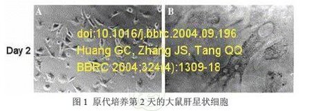
Precautions
2. It is completely feasible to use a solution that does not require in situ digestion, but the digestion time is more difficult to control! The cells in the primary culture or after passage for 9 generations (preferably within 5 generations) were used in the experiment. other
Cell Technology Topic: Primary Culture of Rat Hepatic Stellate Cells
SD rat
Pentobarbital sodium calf serum DMEM enzyme digestive solution D-Hanks' liquid phenol red trypan blue HBSS sodium chloride PBS
Scalpel surgical scissors nylon mesh phase contrast microscope fluorescence microscope
First, the preparation of experimental materials
2. Reagents: sodium pentobarbital, calf serum (or better fetal bovine serum), DMEM, enzyme digestive juice I, II (with HBSS), 18% Nycodenz (with GBSS), D-Hanks 'Liquid (KCl 0.4 g/L, KH 2 PO4 0.06 g/L, NaCl 8.0 g/L, NaHCO 3 0.35 g/L, Na 2 HPO 4 .7H 2 O 0.06 g/L or Na 2 HPO 4 0.053 g/ L, can add phenol red 0.02 g / L), HBSS, trypan blue and so on.
3. Instruments: surgical instruments, 200 mesh nylon mesh, phase contrast microscope, fluorescence microscope.
Second, the primary culture method
1. After abdominal anesthesia in rats, laparotomy on the ultra-clean table, portal vein cannulation, D-Hanks' liquid perfusion, while cutting the inferior vena cava, flushing the blood, and then changing the irrigation with 100 ml enzyme digestion I (0.05 % type IV collagenase).
Third, the method of passage
Fourth, the results
On the next day of inoculation, the cells were star-shaped under light microscope, and there were lipid droplets in the cytoplasm. Autofluorescence was observed at 328 nm under fluoroscopy. Attach a photo published in the Biochem Biophys Res Commun 2004 article (Figure 1).
1. Further identification requires immunohistochemistry: Desmin and α-SMA; the latter is expressed after cell activation.
Expand  First, the reference
1. Yuan Taoxia, Zhang Jinsheng, Zhang Yuexi, et al. In vitro culture of rat liver Ito cells and observation of heparin inhibition. Journal of Shanghai Medical University, 1996; 23(2): 90-93.
2. Huang GC, Zhang JS, Tang QQ. Involvement of C/EBP-α gene in in vitro activation of rat hepatic stellate cells. Biochem Biophys Res Commun 2004;324(4):1309-1318.