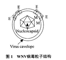
What is the West Nile virus that invades the United States and Europe? 1. Introduction to West Nile Virus West Nile Virus (WNV) was first discovered in Uganda, Africa, in 1937, and was isolated from the blood of a hot adult woman in the West Nile area, hence the name West Nile Virus. West Nile virus belongs to the genus Flavivirus of the Flaviviridae family. The members of the Flaviviridae family include more than 70 species such as dengue fever virus, Japanese encephalitis virus and yellow fever virus, and most of them belong to arboviruses. West Nile virus is widely distributed in Africa, the Middle East and West Asia, mainly causing West Nile Fever. Most of the diseases caused by zoonosis are zoonotic diseases, causing serious public health problems and lack of effective treatment, prevention and prevention. Control measures pose a serious challenge to disease control. 2. West Nile virus etiology West Nile virus is an RNA virus belonging to the family Flaviviridae, yellow virus, and the virus is spherical and has a capsule. The viral ion is about 40 nm, and the viral nucleic acid is a non-segmented single-stranded positive-stranded RNA of about 10,000-11,000 bases. Three structural proteins are encoded: viral capsid protein (C), premembrane protein (preM), envelope protein (E) and seven non-structural proteins: NS1, NS2a, NS2b, NS3, NS4a, NS4b and NS5. E protein is the most important antigenic structural protein of West Nile virus, mediates the binding of virus to host, stimulates the body to produce corresponding antibodies, is the decisive protein that determines the virulence of virus, is a viral hemagglutinin and mediates virus-host binding. . 3. Epidemiology and communication West Nile virus is transmitted mainly through the bird-mosquito-bird, human and animal pathways. The virus can infect humans, birds, a variety of mammals, amphibians and reptiles. People and birds are most susceptible, and the diseases caused by them are mainly distributed in Europe, Africa and the Middle East connected along the bird migration route. country. Its main source of infection is the virulence-bearing animal, and the natural storage host of the virus, the wild bird. The mosquitoes that transmit the virus mainly include Culex, Aedes and Man mosquitoes, especially Culex pipiens. The virus spread to people. Mosquitoes are poisoned by bites that infect WNV birds, and then poisonous mosquitoes transmit West Nile virus to people and other things through bites. West Nile virus can cross the blood-brain barrier and interfere with normal central nervous system function. The clinical manifestation is mainly encephalitis or meningoencephalitis. West Nile virus disease mainly occurs in late summer or early autumn, and WNV can occur in all seasons in warm climates. Organ transplantation and blood transfusion may also be the means of transmission. In 2002, the Georgia Department of Public Health and the US CDC reported two cases of unexplained fever and encephalitis. The two patients were transplanted with the same infection with West Nile virus. Human organs. In addition, breast-feeding and wound invasion are also possible routes of transmission, and attention should be paid to protection. People, animals and birds are all susceptible animals. Such as viruses can infect birds (mainly crows), mosquitoes (Culex, Aedes and Mantis), humans, night monkeys, horses, dogs, cats, pigs, camels, chickens, ducks, geese, pigeons, cattle, bats, Blue birds, squirrels, and rabbits. There have been no cases of West Nile virus infection in China so far, but there are many places in China where the summer is humid and hot, and some places are flooded with conditions for mosquito breeding. In addition, trade and tourism between China and the rest of the world People and personnel are increasingly frequent and have the potential to spread West Nile virus. Therefore, it is very necessary to establish a control technology reserve in China. 4. Clinical features of West Nile virus infection 4.1 Poliomyelitis syndrome It is characterized by high fever above 39 °C, early manifestations of headache, burnout, chills, night sweats, myalgia and confusion. Severe muscle weakness is also a common symptom. Bilateral or unilateral upper limb muscle weakness develops progressively, lower limb weakness Even paralysis; bladder dysfunction, acute respiratory distress. 4.2 West Nile virus encephalitis About 1 / 300 ~ 1 / 150 West Nile virus infection can develop aseptic meningitis, encephalitis or meningoencephalitis, the incubation period is about 2 ~ 14 days, clinically expressed as fever, headache, convulsions, consciousness Encephalitis or meningoencephalitis symptoms such as disorders and meningeal irritation. 4.3 West Nile 80% of people infected with WNV have no clinical symptoms and can recover on their own. The typical symptoms of West Nile virus infection are West Nile fever, which accounts for about 1/3 of infected people. The incubation period is generally 1 to 6 days. Clinically, it shows fever, headache, fatigue, fatigue, lethargy, fatigue, sudden onset of symptoms with or without prodromal symptoms, and fever of 1 / 3 or more patients can reach 38.3 to 40 °C. Some patients may also have severe eye pain, conjunctival edema, congestion and muscle soreness. 4.4 Equine infection Equine animals rarely develop clinical symptoms after infection with WNV, but 10% of cases develop severe neurological diseases, mental depression, loss of appetite, ataxia, muscle tremor, paralysis of hind limbs, unclear vision, Turning around, unable to swallow, excite, lick and even die. 5. Detection method 5.1 Serological testing Serological testing is the most commonly used laboratory clinical test method. The method is an enzyme-linked immunosorbent assay (ELISA) for the detection of West Nile virus IgG and IgM antibodies, in many cases replacing the hemagglutination inhibition test, the complement fixation test and the neutralization test. The most effective one is the ELISA method for capture IgM. The immune response of IgM appears earlier than IgG after WNV infection. The capture ELISA can be used not only to detect IgM of WNV in serum or cerebrospinal fluid, but also has rapid and high sensitivity. Sexual and quantitative, so this technique can be used for early diagnosis of WNV infection, and the sensitivity is higher than PCR detection. This method is the most sensitive screening method for detecting viral antibodies in serum and cerebrospinal fluid. 5.2 Pathogen detection Specimens commonly used for virus isolation include the patient's cerebrospinal fluid, viremia sera (for about 5 days), brain tissue, horse's brain and spinal cord tissue, and the bird's kidney, brain, and heart tissue. It is common to use a known mammalian cell line sensitive to West Nile virus, such as Vero cells or mosquito cell lines, to isolate the virus. Virus isolation is a classic technique for detecting viruses and is the "gold standard" for diagnosing viral infections. It is a very valuable tool for the isolation of new diseases and the natural host of viruses, but this method requires high conditions, low separation rate, and operation. Complex, long time, can not quickly and massively diagnose the virus. In addition, nucleic acid detection methods are also used for the detection of West Nile virus. For example, RT-PCR, Taq Man RT-PCR analysis and nucleic acid amplification (NASBA) can be used to identify whether the virus isolate is West Nile virus. 6. Related test product information Our company provides West Nile virus ELISA test kit, which can be used for the diagnosis of West Nile virus infection. The specific product information is shown in the following table: Product number product name Product specifications Methodology Test sample Brand WNMS-1 Capture Fasinel Nile Virus IgM Test Kit 96T Capture ELISA serum American InBios WNGS-1 West Nile Virus IgG Test Kit 96T ELISA serum American InBios 7. References [1] Lü Wei, Yu Ya, Hu Guixue, Zhao Zhe. Research Progress of West Nile Virus Disease[J].Acta Economica Sinica,2012,16(01):55-59. [2] Fu Yuguang, Ren Qiaoyun, Luo Jianxun, Yin Hong.Research progress in the diagnosis of West Nile virus[J].Chinese Journal of Zoonoses,2011,27(08):742-745. [3] Chang Hua, Hua Qunyi, Xiang Xun, Zeng Zhaowen, Duan Gang. Progress in West Nile Virus Research[J]. Journal of Yunnan Agricultural University, 2006(01): 76-80. [4] Li Yongping, Feng Guanguang, Yi Yuzhen. Research status and prospects of domestic retinoblastoma[J]. Chinese Journal of Ophthalmology, 2004(04): 4-6. [5] Wang Zheng, Wang Linchuan. Overview of Epidemiological Study of West Nile Virus[J].Journal of Guangdong Animal Husbandry and Veterinary Medicine Technology, 2004 (02): 17-18 + 21. [6] Siddharthan V, Wang H, Motter NE, et al. Persistent West Nile Virus associated with a neurological sequela in hamsters identified by motor unit number estimation [J] .Virol, 2009 ,83:4251-4261. Want to know more product information, scan code to pay attention to WeChat public number Amino acids are biologically important organic compounds composed of amine and carboxylic acid functional groups, along with a side-chain specific to each amino acid. With biological significance, amino acids are important in nutrition and are commonly used in nutritional supplements, fertilizers, and food technology. Industrial uses include the production of drugs, biodegradable plastics, and chiral catalysts. Natural Amino Acids,Amino Acids Powder,Amino Acids Particles,Amino Acids Tablets SINOCHEM PHARMACEUTICAL CO., LTD , https://www.sinochemnutrition.com