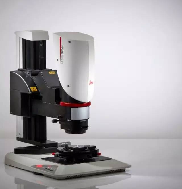
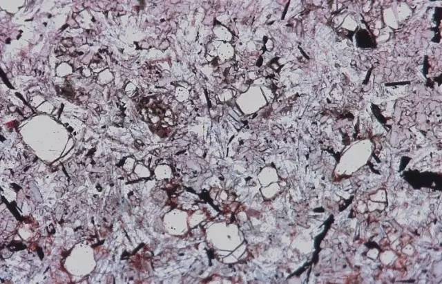
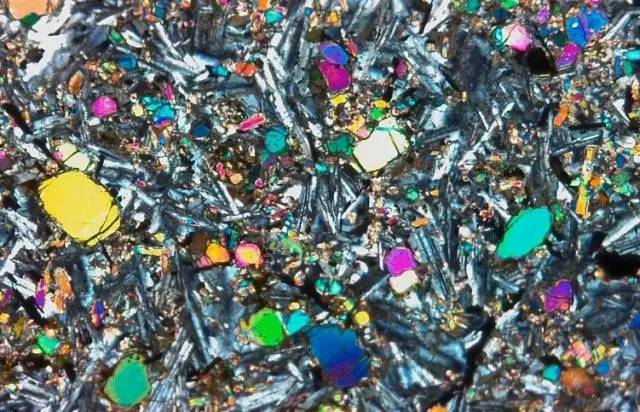
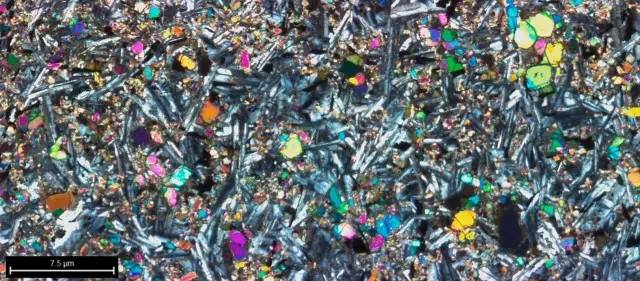
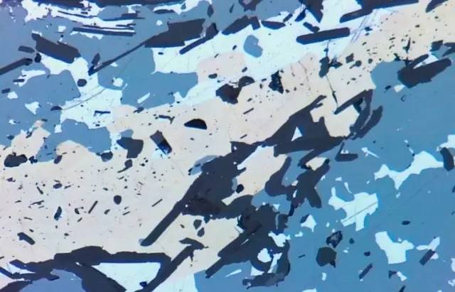
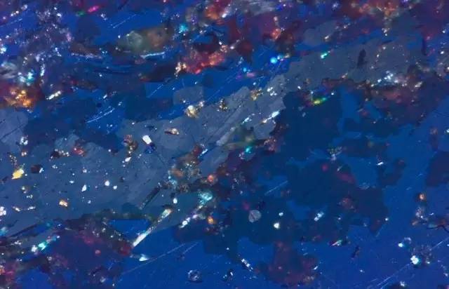
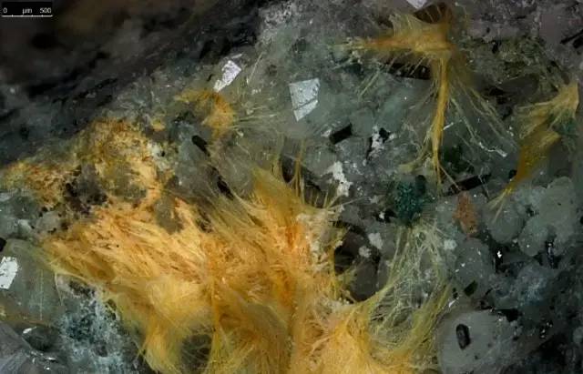
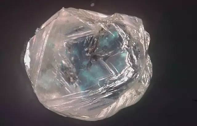
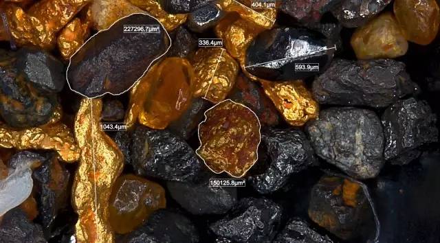
A hundred years ago, polarized light microscopy has been used in traditional earth science research. Since then, with the advancement of technology, such microscopes have gradually improved in terms of user friendliness, ergonomics, and optical performance. Today, there is still one step in place: traditional polarized (multiplex) microscopes are only suitable for prepared samples because they provide a working distance that is insufficient for the entire sample to be tested. This means that thicker, larger geological samples must be cut and polished to accommodate the limited working distance of the compound microscope. These sample preparations require extremely high precision, and the accuracy requirements are even greater in terms of the thickness, flatness, and polishing of the polished sheet. When using a compound microscope with transmission and polarized light [1, 2], the standard thickness should be 30 microns. In other words, when scientists detect unprepared samples, they need to switch to a stereo microscope with a large working distance. Introduction A microscope for testing with or without prepared samples This article describes how geoscientists can analyze samples that have been or are not prepared using a polarized light microscope: this is the Leica DVM6 M digital microscope. With the right accessories, the microscope can be turned into a semi-quantitative polarizing microscope. The diversity of geological samples requires a range of qualified microscope solutions: Suitable for all geological testing projects With the adapter, the Leica DVM 6 M can be mounted on a manual or motorized focus column for additional working distance, ensuring that geologists can view polished or unpolished samples. A high optical aperture transmission mount provides a digital solution for a wide range of polarized applications. A transmissive stage is provided with a polarizing stage with a screw-in/out polarizer. Set up the Leica DVM6 M as described above to ensure that the microscope is suitable for any geological science application and that the user is able to observe transparent samples in parallel and cross-polarized light, respectively. It is not important whether the sample is fitted with a coverslip or whether the sample is uncut clear mineral (for example, diamond or indicator mineral). The Leica DVM6 M is able to increase the productivity of geoscientists due to the following advantages: • Flexible processing of samples with or without preparation using one instrument; • Fully integrated ring light, plus coaxial and other illumination types, ensuring that the options for research and analysis of samples exceed the duplex microscope; • Efficiently and quickly achieve full-scale magnification switching; • Intuitive software interface for microscope operation and analysis; • Has the ability to automatically track and store (encode) important parameters. For more details on the Leica DVM6 digital microscope, see the Leica DVM6 product page [3]. Analysis of prepared samples Take the basalt sheet as an example below. The structure and composition of basalt can be demonstrated by polarized illumination in different directions. Figure 1: Image of basalt sheets acquired using parallel polarized light Figure 2: Image of the same sample taken with crossed polarized light If a single image shows that the region of interest of the sample is larger than the field of view of the microscope, the researchers can use the Live Image Builder feature in the microscope software. During the stage scanning phase, this feature creates a single image from the composite image recorded by the camera, ensuring that the user can view the sample in a wider range. Figure 3: The above picture is a large overview of basalt flakes For reflective, the polished sample can be viewed with built-in coaxial illumination without additional components. In the reflected light mode, the built-in 1â„4 λ plate (wave plate) is adjusted to complete the polarization crossover. Figure 4: Observing ore minerals using parallel polarized reflected light Figure 5: Observing the same sample using cross-polarized reflected light The Leica DVM6 M coaxial illumination mode features a tilted illumination slider that provides a three-dimensional image of grain boundaries, hardness differences and scratches. Simply look at the sample from this angle by simply moving the illumination slider around. No other action is required. The image to the right shows that the Leica DVM6 M with only integrated ring light is suitable for this field. While traditional polarized light microscopes do not have a dark field, the Leica DVM6 M with ring light provides an additional dark field angle for polishing samples. Figure 6: Detail image of the same sample taken with parallel polarized oblique reflected light for viewing hardness differences and scratches Figure 7: Detail image of the same sample taken with ring light illumination Analysis of unprepared samples In addition to these “classic†methods of observing geoscience samples, users can also use the Leica DVM6 M to view three-dimensional images of unprepared samples, such as microscopic slides, core sections, minerals, microfossils, and more. Due to the built-in part of the stereoscopic light arrangement of the stereo microscope or the gooseneck spotlight, it is possible to observe the unprepared sample in a manner similar to a stereo microscope. For samples with a thickness exceeding the depth of field of the microscope, it can be easily observed with the multi-focus function. Subsequently, a three-dimensional topography of the sample can be reconstructed. Also, if you only need 2D images, you can enable the Live Image Builder feature. Microslide samples usually require extended depth of field to make all the details clearer. The Leica DVM6 M software features Live Image Builder Z (with manual focus column) and montage options (custom, EDOF, boot), providing a very simple way to get multi-focus images [3]. Figure 8: Detail image of a microsatellite sample taken with ring light illumination Figure 9: Multifocal image of a microscopic slide sample taken with annular light illumination. Figure 10: Multifocal image of unpolished diamonds obtained with transmitted polarized illumination. Loose samples, rock samples, and cores are usually larger than the field of view provided by the microscope, and Live Image Builder XY can overcome this limitation and combine multiple images into a single image with a larger field of view. The leica DVM6 M has three available objectives with a wide range of magnifications (1x ~ 2, 350x) and resolution, enabling users to quickly switch between large and micro images. Since all images are automatically calibrated, these images can be used for measurements. In 3D topography reconstruction, surface properties and volume can be measured. Figure 11: Multi-focus images of gold and heavy minerals obtained by length and surface measurements. in conclusion This paper demonstrates the possibility of using a microscope to analyze samples that have been or have not been prepared in the field of geological science. The leica DVM6 M digital microscope combines macro and micro to ensure that users can analyze both 2D and 3D samples. Since the field of view and depth of field can be increased at the same time, it is easy to operate samples of different heights. The Leica DVM6 M universal illumination method offers more options than a compound microscope for research and analysis of samples.
Solas Reflective Tape
1) Material of our Solas Reflective Tape :
Our Marine reflective tape for Safety is based on premium polyester material, SOLAS (Safety of Life at Sea) Grade. The surface is equipped with a highly visible honeycomb pattern , with alternating ultra bright silver color, it ensure the reflective safety tape reflect light better in the dark, with smooth and glossy surface that reflects light in any situation added visibility.the high reflective tape gives you extra security.
2) Certificate of our Solas Reflective Tape :
It is a SOLAS (life safety at sea) certification, please note that there is a SOLAS mark every 4cm on the tape.
3) High luminance
our specially made glass microbead tape is manufactured by professional technology. High visibility ensures safety tape reflects light better in the dark, which will greatly increase visibility during the day and at night. Its unique design of glass microbead pattern gives off bright light when reflecting light. Improve visibility, believe he's right, improve SAFETY and reduce the chance and risk of an accident.
4) Features : Industrial adhesive, easy to apply. PET tape has a strong adhesive force, and a certain degree of flexibility, to resist peeling and tearing, strong adhesion. Stickiness can even outlast life. Our reflective safety sign tape is tough, durable, and able to withstand elements for great indoor or outdoor use, and in continuous efforts for safe human travel...
5) Sizes: We have made this reflective tape into different sizes to facilitate your specific needs. Including heat and humidity.
6) Applications: Reflective tape application is very wide, there are many kinds of application such as car parks, warehouses, storage unit, office, classroom, hospital,, garage, roads, factories, machinery, restaurant, goods, garage doors, truck, boat, mailboxes, helmets, different types of trailer, truck, mailbox, bicycle, car, rv, backpacks, railing, ramp, trailers, drivers, hiking, biking and jogging in the daily use, the danger zone. Our tape has infinite possibilities.
Solas Reflective Tape,solas grade reflective tape, Marine Reflective Tape,High Intensity Reflective Tape Kunshan Jieyudeng Intelligent Technology Co., Ltd. , https://www.jerrytape.com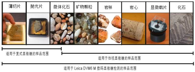


How to analyze a geological sample with or without preparation using a digital microscope
7) How to use: Easy to use. Clean and dry required surface area. Cut the required length of tape, when you stick the tape on the surface, remove the tape and press it into place, be sure to paste successfully once, do not paste repeatedly.