Release date: 2016-02-29 Doctors and engineers at the University of Melbourne in Australia are using supercomputers to create cardiovascular 3D models for patients with heart disease. They sent images collected during routine angiography to a supercomputer. Within a 24-hour period, a 3D model of a human artery was printed in 3D. This gives cardiologists critical information about blood flow behavior and a more precise structure of the arteries in the body. Source: Tiangongshe Bulk Garlic Powder,Bulk Organic Garlic Powder,Minced Garlic Powder,Ried Garlic shandong changrong international trade co.,ltd. , https://www.changronggarliccn.com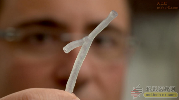
Scientists also said that it can also help doctors choose the most appropriate stent for their patients (a device used to open and keep the artery folded or blocked). In addition, the technology is able to detect plaque hotspots that accumulate in the arteries and cause heart disease. Some plaques are difficult to find using traditional techniques.
Scientists have now published their research in the academic journal "European Heart Journal" published on February 23, 2016.
To date, heart disease remains the number one killer in Australia, affecting one in six adults in the country. According to statistics, there is a heart attack every 9 minutes in Australia. Researchers estimate that this new technology that can detect plaque in the heart will help reduce the incidence of heart disease in the country.
The first author of the study was Peter Barlis, an associate professor at the University of Melbourne, who is also an interventional cardiologist at t Vincent's and Northern Hospital. 
“Using our ultra-sensitive cardiac scanning technology combined with a supercomputer-derived model, we are now able to print out the patient's arteries in 3D so that we can tailor the matching (bracket) device.†Barlis said: “We It is hoped that using these models will predict the type of stent that best fits the patient. Once these techniques are simplified, our ideal scenario is that a modeled and 3D printed artery can guide the procedure while the patient is lying on the operating table."
But for cardiology, the Holy Grail in the minds of scientists is still determining how plaques can cause heart attacks.
“Using an ultra-high resolution camera called the Optical Coherence Tomography (OCT) to scan the inside of the heart artery has made it easier to image cholesterol plaques, but it is unclear which plaques will cause the heart. Symptoms. If these high-risk plaques are identified more accurately and earlier, then we may be able to prevent them before a heart attack." 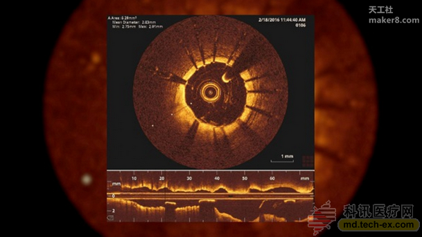
Associate Professor Barlis has been improving the technology that can benefit heart patients since he introduced OCT to Australia in October 2009. He said that 3D modeling has great potential to predict where plaques will form, which will ultimately help cardiologists predict heart attacks.
Dr. Vikas Thondapu, a collaborator of the paper and a researcher at the University of Melbourne, said that clues about dangerous cholesterol plaques may be present in certain factors that interfere with blood flow patterns.
"Our work involves the use of supercomputers to simulate blood flow in arteries. The goal is to predict the future development of high-risk plaques through blood flow patterns and their interference," Dr. Thondapu said.
Associate Professor Barlis and his team have received two research grants and are working with the School of Engineering to find a biocompatible material to 3D print a heart stent that precisely matches a person's anatomy, thereby reducing stent collapse. Or the risk of complications. In addition, they are also interested in new polymers that allow the stent to slowly dissolve over time and deliver the drug directly to the location of the plaque.
Imperial College London and Harvard University are collaborating with the University of Melbourne on this groundbreaking study.